REM Sleep: Definition, Functions, Disorders, Research
Was this post helpful? Let us know if you liked the post. That’s the only way we can improve. Yes 2 No 0
Was this post helpful? Let us know if you liked the post. That’s the only way we can improve. Yes 2 No 0

Carlos is a neuroscientist and a medical & science writer with more than eight years of research experience in the field of Neuroscience. Prior to working full time as a medical writer, he was a postdoctoral researcher at the University Hospital of Bern (Switzerland). Carlos obtained his PhD from the University of Iowa (USA), supported by the Fulbright Program.
Some of the areas Carlos focuses on are RNA therapeutics, Rare Diseases, and REMS/RMPs. He has authored multiple original research papers in top journals in the field, book chapters, and periodicals. Carlos has also participated in international scientific meetings; most notably, he was invited to present his dissertation research at the 2018 Gordon Research Conference on Sleep Regulation and Function.
Sleep is one of those things where the beautiful complexity of our brains come to full shine. There are a lot of things we don’t understand about sleep, but it is clear to everyone that it is a biological necessity.
We need sleep to recover both mentally and physically, integrate memories, learn, stay focused, process emotions, among others. And the more scientists learn about how our nightly rest affects other aspects of our lives, the more they attempt to elucidate some of its mysteries.
That’s how researchers ended up characterizing the sleep cycle and learning about the different sleep stages. In clinical settings, we differentiate light, deep, and REM sleep stages based primarily on different patterns of brain activity, but also in relation to heart rate variability, breathing patterns, and muscle activation.
REM sleep (also called paradoxical sleep, or active sleep) has been the subject of intensive research and discussions since it was first described in the early 50s. The fact that most of our dreaming activity occurs during this sleep stage has also created a mysterious aura around REM in popular culture.
The research on REM sleep is extensive, and it has been going on for more than 65 years. If you are looking to learn more about REM sleep and related phenomena, this article has all the answers.
Sleep wasn’t that exciting among researchers before the 20th century. Until then, sleep was thought to be an inactive state when our brain shuts down due to a lack of response to external stimuli. This understanding of sleep as a passive state made the topic unattractive to most researchers, and it wasn’t until the discovery of REM sleep that they began to pick an increased interest.
In 1953, a team of researchers from the University of Chicago noticed periods of ‘active’ sleep with regular, rapid eye movements (or REMs).[1] It looked like these active sleep periods alternated with periods of quiescent sleep. A few years later, they showed that this state correlates with specific brainwave patterns and dreaming. [2.3]
Besides rapid eye movements, we now know that REM sleep is also characterized by the increased frequency and decreased amplitude of cortical EEG, high-amplitude theta waves in the hippocampal region, muscle twitches, inactivation of skeletal muscles, body temperature fluctuations, autonomic respiratory activation, and increased arousal threshold.[4] Since the EEG readings resemble that of a waking state, but the muscles are shut off, some scientists refer to this state as ‘paradoxical’ sleep. [5,6]
REM sleep is not exclusive to humans. Studies found that most mammals, birds, reptiles, and surprisingly some invertebrates like cuttlefish could experience REM or REM-like sleep.[7,8,9,10] And even though it was first believed that REM is connected to homeothermy (i.e., the ability to keep a stable internal temperature) since it was only observed in mammals and birds, recent research clearly showed that some reptiles exhibit some key features of REM, including rapid eye movements. That means that the origin and evolution of REM sleep probably go way back before the era of birds and mammals, and could be present in all amniotes. Although these animal groups share a common ancestor, the duration and characteristics of the different sleep stages are quite different between species, suggesting that sleep has been shaped by ecology and evolution [11] For instance, sleeping mice experience REM sleep every 10-15 minutes, while in humans, it occurs every 90-120 minutes.[12.13] And the duration varies across species as well; while we spend around 25% of total sleep time in the REM phase, giraffes, for instance, spend less than 5%. [14]
REM sleep can be accurately detected and analyzed in a controlled environment, like a lab or sleep center. In a sleep study, subjects are attached to a series of electrodes and other medical devices that measure different attributes related to sleep, namely:
Body temperature, respiration, and arousal threshold can also be measured in these controlled conditions. This whole process of measuring sleep is called polysomnography and is usually performed to detect the cause of sleep problems in people struggling to get enough rest.
Sleep studies on animals include similar measurements and are mostly performed in rodents and other species in captivity.
Animal studies have provided interesting insights into the mechanisms that originate and sustain REM.
The brainstem is a part of our central nervous system (CNS) responsible for some of the primary physiological functions like breathing, heartbeat, swallowing, blood pressure, and sleep and arousal. Because of that, it was a natural candidate to inspect and find out whether it has any control over some of the components of REM sleep.
The early studies on cats identified part of the brainstem called the pons to be responsible for muscle atonia during REM sleep.[15] Researchers found that lesions in this region caused animals to exhibit overt motor behavior during REM since their skeletal muscle activity was disinhibited. Later studies located the exact pontic region responsible for this, the so-called sublaterodorsal tegmental nucleus (SLD). [16]
SLD and caudal laterodorsal tegmental (cLDT) neurons produce glutamate, which is a common excitatory CNS neurotransmitter. They are mostly active prior to and during REM sleep, and by activating inhibitory interneurons in the ventral medulla and spinal cord, they produce muscle atonia.[17] Disrupting the activity of these neurons leads to REM without atonia. [18,19] On the other hand, several studies have shown that manipulating different neuronal subgroups in these brainstem structures can affect other aspects of REM sleep, including REM quantity and cortical activation.
We also know that neurons in the lateral hypothalamus that secrete melanin-concentrating hormone (MCH) are particularly active during REM sleep.[22] They could possibly promote REM by inhibiting neurons in other midbrain and brainstem regions. Neurons in supramammillary hypothalamus are also essential for REM sleep control and theta wave activity, additionally highlighting the complexity of network circuits in REM regulation.[23] In summary, brainstem, midbrain, and hypothalamus remain the most crucial brain parts when it comes to REM sleep.
Do genes play any role in the regulation of REM sleep? Although this is not an easy question to answer, scientists have made substantial efforts to understand the genetics of REM sleep
A 2015 study investigated receptor coding genes for one of the most abundant neurotransmitters – acetylcholine.[24] This neurotransmitter is essential for neural signaling, and increased levels during sleep are detected in the cortex and hippocampus. Chrm1 and Chrm3 are two of the five acetylcholine receptors suspected to play a role in REM sleep. Using genetic tools, researchers silenced the genes coding for these receptors in mice and then observed the effects it had on sleep. Interestingly, this silencing led to a massive decrease in REM and non-REM sleep. Mice lacking Chrm1 showed fragmented and shorter REM, while Chrm3 inactivation also affected non-REM sleep. When both genes were shut off, it led to almost complete elimination of REM.
These findings illustrate how genetic tools can give us exciting insights into REM sleep. Understanding how genes regulate sleep could help us better understand how our bodies generate sleep and the etiology of sleep disorders.
We know sleep is essential for proper functioning, but can we say the same about the REM phase? Would anything change if REM sleep didn’t occur, or is it crucial for us as any other part of the sleep cycle?
It turns out that REM is vital for many aspects of our lives. Therefore, we split this section into a few different categories to organize all the interesting details you need to know about the functions of REM sleep.
Early studies concluded that newborns sleep way more than adults, and that showed to be the case for both humans and other mammals. Interestingly, the relative amount of REM and non-REM sleep in early development is also different from adults. The graph below depicts how the relative time spent on REM sleep changes across the lifespan in humans.
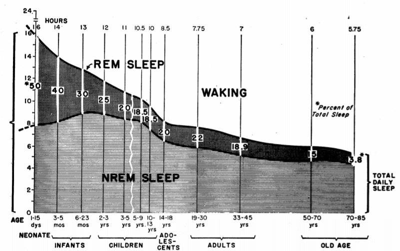
Source: Roffwarg, HP, Muzio, JN, Dement, WC. (1966). Ontogenetic development of the Human Sleep Dream Cycle. Science
Newborns snooze for 16 hours a day, and they spend around 50% of that time in REM. This percentage decreases with age, and adults spend less than 25% of their time asleep in REM. So, why is this happening?
A 1966 article published in the journal Science proposed an explanation for this relative increased REM in the early stages of development.[25] It turns out that neural activity is essential for the rapid growth of the nervous system. The authors argued that, since babies spend most of their time sleeping, they don’t receive enough external stimulation to keep their brains active. Remember that REM is also called “active sleep”? Indeed, during REM sleep, the eyes move rapidly, muscle twitches are seen throughout the body, neural circuits get activated… and all this activity supports the development and shape the young body and mind. Thus, according to this “ontogenetic hypothesis,” REM sleep provides necessary activity for brain circuits to develop. This notion would also explain why the proportion of REM sleep decreases with age. As infants get older, they have more and more opportunities to interact with the outside world and receive lots of stimulation during wakefulness.
Similarly, it seems that the abundant muscle twitches that babies experience during REM sleep can stimulate sensorimotor development in the early stages of life. If you are wondering how we go from crude limb movements in infancy to fine motor control in adulthood, be aware that twitching during REM may play a significant role there. Scientists have found out that sleep twitches evoke sensory responses in the newborn brain, and thus stimulate neurons throughout the nervous system. Because babies spend a lot of time in REM sleep, and because twitches are so abundant during this sleep stage, scientists also believe that they contribute to the wiring of new neuronal connections in the sensorimotor system.[26] Additionally, myoclonic twitches during active sleep have been shown to trigger and synchronize neural oscillations across brain areas, potentially contributing to synapse formation, neural differentiation and migration, and neural connectivity. [27]
Another REM function important for development is the strengthening and maintenance of newly formed connections between neurons, or synapses.[28] A key feature of neural development is the formation of abundant, excess synapses; however, only a small number of those will be maintained through life. The removal of these excess connections is called “synaptic pruning,” and it was recently shown in young mice that REM sleep deprivation disrupts this process.
That last segment already hinted at how REM sleep could be responsible for our ability to store memories and learn new things. Synaptic pruning and strengthening is not only essential during development, but it is also needed for learning. Synaptic connections are vital for memory forming, but only a small portion of newly formed circuits are maintained. Essentially, the brain chooses which circuits to keep, and it incorporates them into a vast neural network of existing knowledge and memories. Over 75% of new synapses get pruned right away while the rest are kept, which could lead to the formation and incorporation of new memories. [30] And again, it looks like active sleep is when all of this happens.
In 2016, scientists managed to highlight neural oscillations during REM sleep responsible for memory consolidation. [59] They showed that silencing specific neurons during REM phase impaired newly obtained contextual memory, while doing it at any other point had no such effect.
However, remember that not all memories are the same. Memorizing random facts activates different brain centers than learning how to ride a bicycle, or remembering a highly emotional event in your life. It seems that REM sleep doesn’t play the same role in every type of learning, and its role in memory consolidation is still controversial. However, REM sleep can “rescue” memories after they have been heavily damaged by interference, which typically occurs when other memories prevent retrieval of certain information. [32]
Studies in both humans and animals show that REM sleep plays a significant part in procedural, spatial, and emotional memory.[33,34,35,36] Although there are inconsistencies across studies, this suggests that REM may not be involved in the consolidation of declarative memories (remembering random facts), which is thought to be supported by slow-wave sleep. Many of the studies showcasing the benefits of REM on memory and learning showed that post-learning REM boosts performance in cognitive tasks. Typically, control groups stayed awake or slept after the learning process, but were prevented from entering REM sleep. While the conclusions of these studies are valid, researchers failed to see such a link in many other experiments. [37,38]
One of the biggest arguments against the role of active sleep in memory consolidation is that the techniques used to suppress it often causes a certain amount of stress, and the learning impairment could be the result of that.[39] There have been reports of patients with lesions in the pontic area, which is essential for REM generation, and these individuals didn’t experience paradoxical sleep. It is interesting that these patients led normal lives and had no problem with memory consolidation.[40]
To make things a bit more complicated, another study where researchers investigated a link between active sleep and memory retention showed interesting results. Pharmacologically induced REM suppression didn’t impair skill learning; in fact, it enhanced it.[41] The results of these studies are sufficient to make us question the true nature of REM sleep. More studies are needed to draw any firm conclusions.
Some scientists concentrate on the physiological functions of REM sleep. For instance, according to the energy relocation hypothesis, organisms evolved three different strategies to maximize the efficiency of energy usage. One of them includes sleep-wake rhythms, where animals would experience periods of rest associated with less expenditure of energy so the organism can focus on other biological processes compared to wakefulness. [42]
It’s not that sleeping saves energy for when we are awake, it’s that it is spent differently. For instance, when awake, animals need to mate and find food, shelter, and other resources, which limits their ability to perform different functions like the growth of neurons, myelin formation, and other brain functions. This is where sleep comes in! As for REM sleep specifically, suppression of both thermoregulation and muscle tone saves a lot of energy, which can then be used to maintain sleep-dependent CNS physiological functions.
This hypothesis makes perfect sense when we are talking about endothermic or “warm-blooded” organisms, like birds and mammals. These animals are able to control their optimal body temperature via internal means. Because this internal regulation of temperature consumes a lot of energy and is specifically suppressed during REM sleep, the energy allocation hypothesis argues that REM has evolved to, so to speak, give the body a break so it can focus on other biological needs. However, with the discovery that reptiles and possibly other animals could be able to experience REM sleep, we are not sure where that would fit in the energy relocation hypothesis.
A 2019 study showed that sleeping mice who experienced warm ambient temperature within their thermoneutral zone (i.e., the range of ambient temperature where the body can maintain its internal temperature without needing to use more energy than the basal rate) increased their REM sleep duration, as compared with mice who in the cool or constant baseline ambient temperature conditions.[43] These findings support the energy allocation hypothesis, as mice opportunistically expressed more REM sleep in the warmer environment, where they didn’t have to spend that much energy in regulating their internal temperature
REM sleep also affects hormone production and metabolism. For example, REM sleep deprivation disrupts leptin homeostasis, which is a hormone that plays a massive role in modulating appetite. That could be one of the reasons why many studies found a link between lack of REM and sleep in general, and weight gains, and obesity. [45]
Studies of abnormal REM sleep control primarily focuses on identifying neural circuit mechanisms leading to these disorders. Interestingly, narcolepsy and REM sleep behavior disorder show many similarities in the deterioration of mechanisms leading to healthy REM sleep.
Rem Sleep Behavior Disorder (RBD) is a neurological condition where patients lose their ability to suppress muscle tone during active sleep. [46] Symptoms vary from exaggerated muscle twitching to shouting, sudden violent movements of limbs, to complex behaviors where people look like they are trying to reenact their dreams.[47] Although it may not seem like a lot, individuals with RBD can injure themselves or their partner during sleep.
But the most worrying thing about RBD is that the majority of patients develop a neurological condition 6-15 years after the onset of RBD symptoms.[49] Most people develop Parkinson’s disease, multiple system atrophy, dementia with Lewy bodies, and other synucleinopathies due to neurodegeneration.[50] This link indicates that RBD manifests due to neurodegeneration, and the new evidence points out to deterioration of neurons in the brainstem, precisely dorsal pons. [51]
Considering that we already found these centers to be responsible for producing REM atonia and playing an active role in other parameters of paradoxical sleep, it is clear why degeneration of these areas leads to RBD. But, since so many people with RBD develop a synucleinopathy, scientists pose a question whether it can be considered as a separate disorder, or whether we should treat it as one of the symptoms that preclude neurodegenerative diseases. One study found that over 80% of patients with RBD developed a neurological condition, and the survival rates 14 years after the initial diagnosis were only 7.5%. [52]
Synucleinopathies are named after a misfolded protein called alpha-synuclein, which causes the degeneration of neurons. According to Parkinson’s disease pathobiology, this protein originates in the gut and then slowly progresses around the body, eventually affecting the brain. [53] Once they reach the brain, the brainstem is the first place of contact, close to REM circuits. Afterward, it spreads to the other brain structures, causing motor and cognitive impairments that characterize Parkinson’s disease. [54]
Interestingly, while brainstem is an area where many vital functions like breathing, chewing, and swallowing originate, they are unaffected by alpha-synuclein degeneration in RBD. Understanding why these circuits remain untouched while REM ones are widely affected is important for future understanding of these conditions, and possible prevention of RBD.
Narcolepsy is a sleep disorder estimated to affect 1 in 2000 people. [55] It is mostly caused by the lack of neurons that produce orexin in the lateral hypothalamus.[56] This neuropeptide plays an essential role in controlling sleep, wakefulness, and appetite. The most common symptoms include excessive daytime sleepiness, fatigue, irresistible sleep attacks, sleep paralysis with vivid hallucinations, and cataplexy.[57] Cataplexy is described as sudden and transient muscle weakness and loss of muscle tone during conscious wakefulness.
Cataplexy resembles muscle atonia of REM sleep, which is why scientists believe that the same neural circuits are responsible for both. And there seems to be more and more evidence supporting this hypothesis. For instance, certain antidepressants used to suppress cataplexy, also affect REM sleep.[58.59] There is rebound in REM and cataplexy after the withdrawal of medications, pointing to a closely related neural regulation of these events.
Cataplexy is usually triggered by strong emotions like fear, surprise, and laughter. Amygdala is a brain structure involved in processing emotions, and recent studies show it is also active during both active sleep and cataplexy. [61,62]
We already mentioned that RBD precludes a variety of neurological disorders. In addition, it looks like RBD and narcolepsy are closely related, as one study showed that over 50% of narcolepsy patients also experienced RBD.[63] What’s even worse is that many patients who take antidepressants to ease narcolepsy symptoms can also face an increase in REM phase movements, even if they don’t fully develop RBD.
You get the idea of how sudden irresistible cataplexy events in narcoleptic patients can impair even the easiest daily activities. Cooking a meal can turn into a disaster, and narcolepsy patients need to be extremely careful when driving, as you don’t want to be behind the wheel when cataplexy strikes. That is why it is essential for people experiencing narcolepsy symptoms to talk to their doctor immediately. They can run tests, find out what is really causing their sleep problems, and provide advice on how to move forward.
Dreams have captivated us since the dawn of time. From ancient people believing these nightly visions showed the future, artists who claimed their inspiration came to them in sleep, philosophers trying to find the meaning behind dreams, to psychologists considering them to be the door to our unconscious mind, and trying to find out what is really going on behind all of this along with other scientists. However, dreaming remains a mystery to this day.
Dreams can occur during different sleep stages but are most closely associated with paradoxical sleep. Many scientists have given their view on why dreams occur. However, we need more research to draw solid conclusions. Any of the proposed hypotheses could be valid, and they are not even mutually exclusive, as dreaming may serve different functions.
By the continuity hypothesis, your dreaming state reflects conscious experiences and is widely affected by them.[64] So, the content of your dreams is impacted by waking life, emotional involvement, personality traits, and the time of the night when dreams occur. A 2018 study found that people with symptoms of depression and anxiety experienced more dreams with negative and harmful connotations.[65] These findings support the hypothesis that waking life plays a major impact on what goes on in our heads during the nightly slumber.
Some have proposed that dreams help us consolidate memories by playing over events from our life. However, we all know that these events often don’t make too much sense, and while many of us would probably love to be able to fly or teleport like in dreams, it is not very likely. But these nightly visions could help us process emotions, or encounter situations that would make it easier to face them in real life. [44]
Since recollecting dreams is not so easy, scientists are trying to visualize them by tracking brain activity in dreaming subjects. However, it is not an easy task since there is not a one-to-one correlation between activity patterns and dream content; furthermore, even the same dream wouldn’t have the same pattern of brain activity in different people. One study used machine learning models to predict the content of dreams, and it was actually successful.[66] That gives us hope that decoding what goes on in our head during sleep is not such an impossible task.
However, some speculate that dreams may not have to serve any particular purpose and that they are simply a byproduct of our brain activity during sleep. Since neurons fire quite a lot during sleep, dreams could be just a random manifestation of that. Unfortunately, that takes away from the mystery and intrigue surrounding sleep. One thing is sure, we won’t stop being fascinated by dreams, and scientists are trying their best to understand them a little better.
Some random facts about active sleep can paint a good picture of how fascinating this state is. So, let’s look at some of them:
Even though the fascination with dreams has been going on for a long time, sleep science is a relatively new field. In the last few decades, we have learned a lot about what happens during sleep and how it impacts our brain and body. However, a lot of things remain a mystery. So, let’s take a look at what is happening with REM sleep research at the moment.
As you can see, research in this field is very active, and while there are a lot of questions we need to answer when it comes to REM sleep, scientists are doing their best to uncover all the mysteries of this phase, and sleep in general.
Dusan is a biologist, a science enthusiast and a huge nature lover. He loves to keep up to date with all the new research and write accurate science-based articles. When he’s not writing or reading, you can find him in the kitchen, trying out new delicious recipes; out in the wild, enjoying the nature or sleeping in his bed.